Radiation Shielding Material for Medical Treatment
- Categories:Tungsten-molybdenum for Radiation Shield
- Author:
- Origin:
- Time of issue:2017-11-21 00:00
- Views:
(Summary description)I. X-ray pipe side collimator also called front collimator is mounted in front of a CT bulb pipe, and is the first part through which X rays pass. X rays emitting out of bulb pipes are conical, and can obtain a thin light beam [1] after passing through a tungsten device with narrow seams. Its main function is layer thickness selection of X rays. It can control the width of X ray beams in the parallel direction of long axis of a human body, thus controlling the thickness of scanning layers.
Radiation Shielding Material for Medical Treatment
(Summary description)I. X-ray pipe side collimator also called front collimator is mounted in front of a CT bulb pipe, and is the first part through which X rays pass. X rays emitting out of bulb pipes are conical, and can obtain a thin light beam [1] after passing through a tungsten device with narrow seams. Its main function is layer thickness selection of X rays. It can control the width of X ray beams in the parallel direction of long axis of a human body, thus controlling the thickness of scanning layers.
- Categories:Tungsten-molybdenum for Radiation Shield
- Author:
- Origin:
- Time of issue:2017-11-21 00:00
- Views:
Business Consulting:+86-13526941255 Yanyan
Email:yanyan@achemetal.net
High-precision Tungsten Device for CT Collimator and Detector
Collimators are divided into two kinds in medical CT:
I. X-ray pipe side collimator also called front collimator is mounted in front of a CT bulb pipe, and is the first part through which X rays pass. X rays emitting out of bulb pipes are conical, and can obtain a thin light beam [1] after passing through a tungsten device with narrow seams. Its main function is layer thickness selection of X rays. It can control the width of X ray beams in the parallel direction of long axis of a human body, thus controlling the thickness of scanning layers.
II. Detector side collimator is also called rear collimator of which narrow seams are respectively aligned with each detector, so that detectors can only receive rays emitting into detectors vertically, so as to reduce the interference of scattered rays from other directions as much as possible.
Detectors are devices for transforming X-ray signals attenuated after absorption of human body tissues into electrical signals, and can be divided into fixed detectors and gas detectors both of which are selected in modern CT devices. Choosing the category of measurement systems should refer to stressed characteristics. Gas detectors are used for measuring the strength of incident X rays by measuring the magnitude of current when gases are ionized by incident X rays according to the principle of ionization of xenon (Xe), an inert gas. Both partition plates and collecting electrodes of gas detectors are tungsten devices. Being consistent with the incidence direction of X rays, partition plates can achieve the effects of rear collimators, and can prevent scattered rays generated by human bodies to be measured from entering ionization chambers. When on X ray beam enters a high-pressure xenon cavity, xenon will be ionized, positive ions move towards the cathode while negative ions are collected by a collecting electrode (tungsten device), to generate signal current. These signals will directly reflect the X-ray absorption amount of detectors.
Developing rapidly, medical CT technologies have developed to the sixth generation at present. The quantity of detectors is increased further, so the using amount of high-precision tungsten devices is increased obviously, and the devices need replacing at regular intervals.

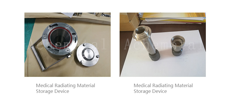
Medical Protective Storage Tank
With a good radiation shielding property, tungsten and tungsten alloy are used for manufacturing radioactive substance shielding walls as well as containers for storing and transferring radioactive substances in the medical field.
 +8613526941255

 Message